


PLAX (Parasternal Long Axis)
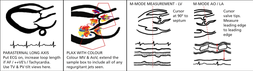
Image from BSE website
2nd to 5th intercostal space just next to sternum. Probe marker pointed towards patient’s R shoulder. The higher the level the more likely the LV will be at 90 for M-mode measurements.
2D
Capture a deep clip looking for pleural and pericardial effusions. 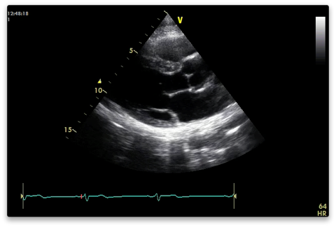
Then:
Narrow sector width and reduce depth to focus on area of interest.
Capture a shallower clip with the heart filling the screen. You are not meant to see the apex of the LV.
RVOT uppermost
Aortic valve - right coronary cusp above; non below
Mitral valve - A2 (upper) and P2 (lower) segments visible
Descending aorta behind posterior MV annulus.
Have a general look around for abnormalities:
- LV function
- Pericardial effusion
- Valves for thickness, mobility and calcification.
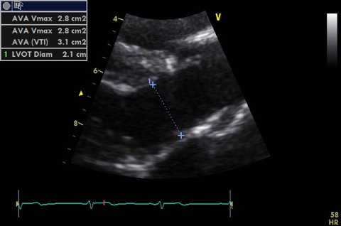
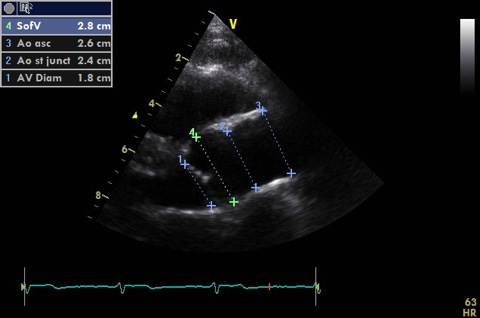
Measure in 2D:
- LVOT (up to 1 cm from aortic annulus - must PW at same point in A5C). Image should be shallow, narrow and zoomed to improve accuracy. Cross sectional area is calculated from this.
- Aortic annulus
- Sinuses of valsalva (widest bit)
- Sinotubular junction (where S of V become ascending aorta)
- Ascending aorta (include a measurement with probe tilted slightly superiorly)
- Proximal RVOT in diastole (in line with AV leaflets). Not shown
- LA end systole (in line with AV leaflets) if not measured with M-mode. Not shown
- LV measurements can be measured in 2D but M-mode preferable (better resolution) if scan line at 90 degrees.
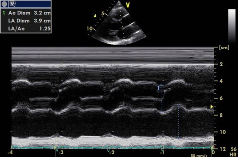
M-mode
Through tips of aortic valve leaflets
- Assess valve opening
- Measure aorta (end diastole) and LA (end systole)
Through tips of mitral valve leaflets
- E point (valve tip at most anterior position in early diastole)
- Measure distance from E point to IVS (EPSS)
- A point where valve opens up again in late diastole with atrial systole

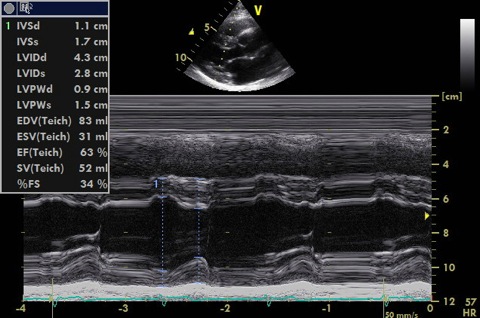
Just beyond tips of MV leaflets at 90 degrees to long axis of LV
- Measure septum, LV cavity, posterior wall in systole and diastole
- These can be measured from 2D if the ventricle is not at 90 to your scan line (which is frequently the case)
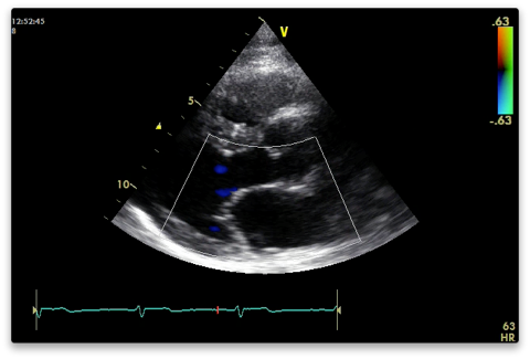
Colour (Nyquist limit 50-60 cm/s)
Aortic and mitral valves for stenosis and regurg. Do one at a time. The resolution is better the narrower and shallower you have the colour box (cutting off the near field makes no difference).
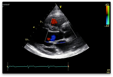
Measure vena contracta if present (narrowest width of regurg colour).
Colour M-mode for AR if present - measure jet width and calculate as percentage of LVOT width.
LVOT for flow acceleration if septal hypertrophy
IV septum
PRVI (parasternal RV inflow)
From the PLAX view tilt the probe anteriorly looking towards the patients right hip.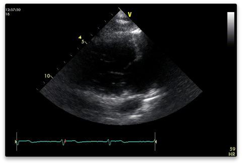
2D
Capture
Anterior (upper) and posterior (lower) tricuspid valve leaflets.
RA, RV, IVC +/-coronary sinus also visible.
Colour
TV inflow and regurg.
Doppler
CW doppler through valve.
If TR measure peak velocity - align with jet.
PRVO
From the PLAX view tilt the probe towards the patients left shoulder.
2D
Capture
RVOT, pulmonary valve and pulmonary artery visible.
Look for thrombus (PE)
Colour
PV - stenosis or regurg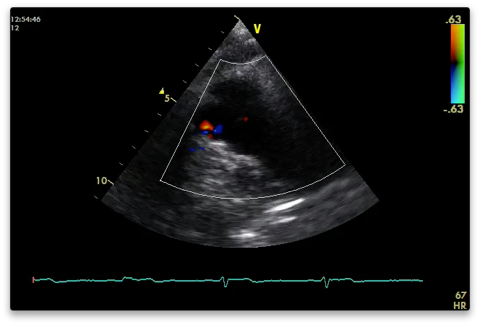
Doppler
CW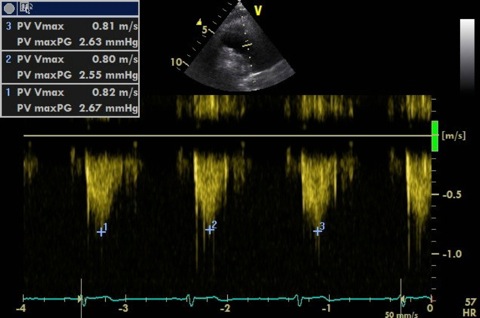
- Measure peak velocity of forward flow for stenosis and trace VTI for mean gradient (BSE recommends PW PV inflow for mean gradient).
- Align with regurgitant jet to measure peak velocity (used to calculate PADP). PR Vmax end diastole for end PADP and PR Vmax early diastole for mean PADP.